• 16
    Feb
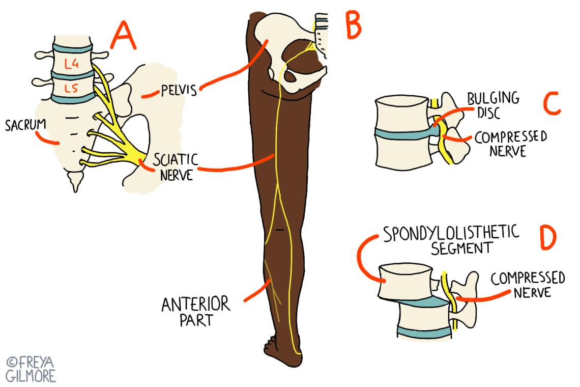
    Diagrams illustrating sciatic nerve anatomy and compression causes. Part A shows sacrum, L4/L5 vertebrae, pelvis, sciatic nerve. Part B depicts leg nerve path. Part C shows bulging disc compressing nerve. Part D highlights spondylolisthesis causing compression. Labeled "FREYA GILMORE."

    Mythbusting in Osteopathy

    Mythbusting in Osteopathy We hear a lot of misinformation in clinic, whether our patients pick it [...]

  • 9
    Feb
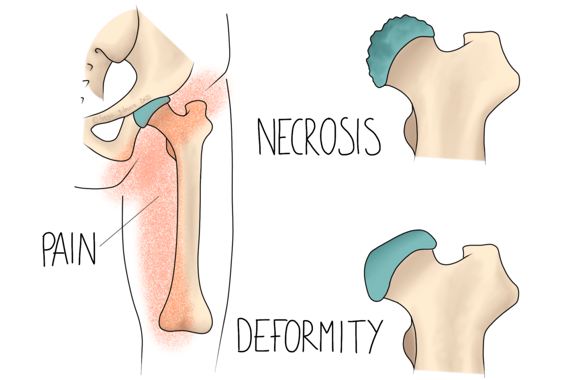
    Hip bone illustration shows necrosis and deformity with highlighted areas indicating pain. Labels: "PAIN," "NECROSIS," "DEFORMITY." Surrounding text and graphics emphasize joint issues.

    Perthes’ Disease

    Perthes' Disease Perthes' Disease is a rare childhood disease affecting the shape of the hip joint. [...]

  • 2
    Feb
    Virus-like illustration displaying symptoms: "muscle & body aches, PROFOUND FATIGUE, weeks - months." Central circular design with purple appendages, text within.

    Long Covid

    Long Covid Long Covid is a poorly understood condition, and new information is still emerging. This [...]

  • 26
    Jan
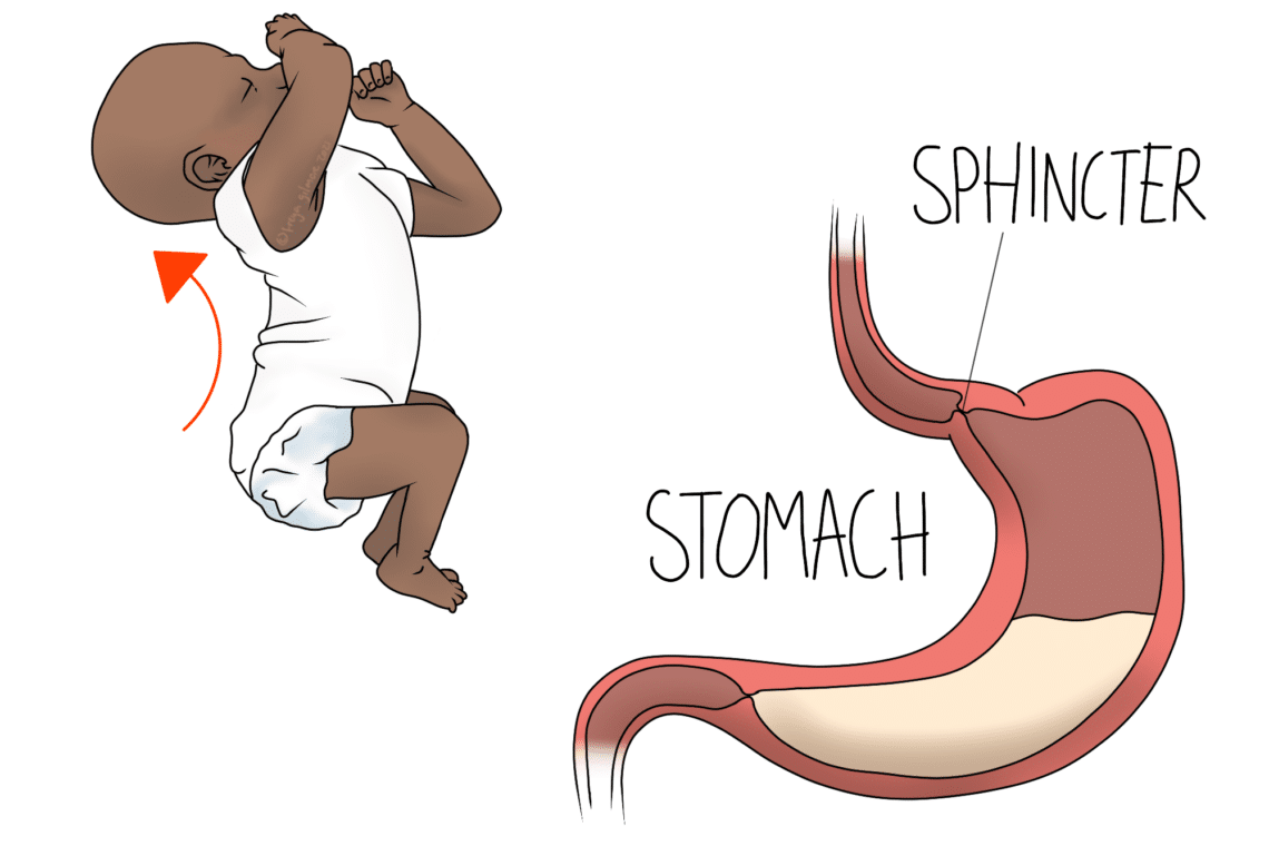
    A baby wearing a white onesie is shown curling up with an upward arrow beside it. Next to this, a labeled diagram illustrates a stomach with "SPHINCTER" and "STOMACH."

    Reflux

    Reflux Acid reflux or heartburn can affect all ages. In this post we will cover both [...]

  • 19
    Jan
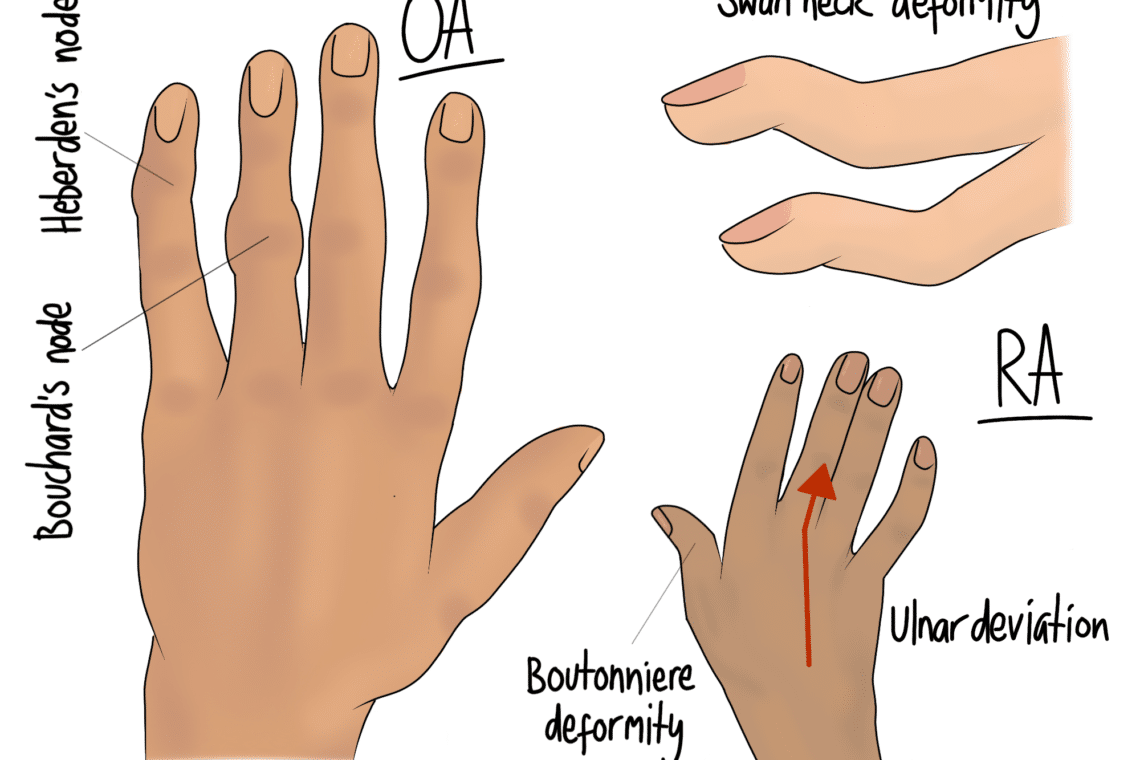
    Two hands depicting arthritis-related deformities. The left hand shows "OA" with Heberden's and Bouchard's nodes. The right hand shows "RA" with swan neck deformity, Boutonniere deformity, and ulnar deviation.

    Rheumatoid Arthritis

    Rheumatoid Arthritis Sometimes patients will mention that they have a family history of arthritis, but they’re [...]

  • 12
    Jan
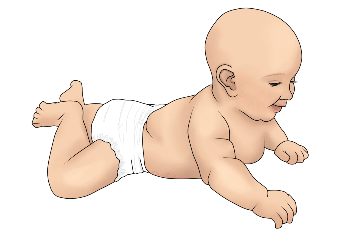
    A baby wearing a diaper is crawling on all fours, appearing joyful in an isolated white background.

    Infant Milestones

    Infant Milestones Healthcare professionals use milestones to help monitor your baby's development. It is important to [...]

  • 5
    Jan
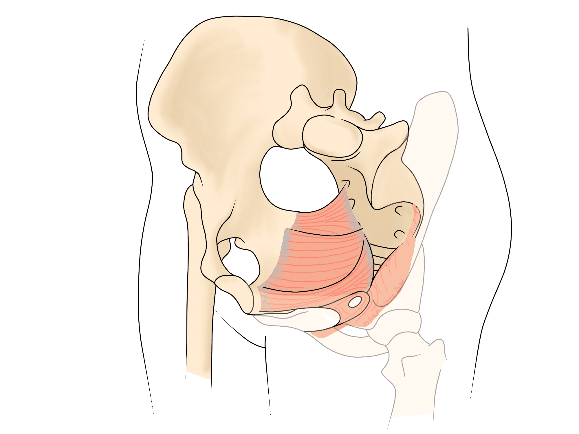
    Anatomical illustration of pelvic bones showing layers of red muscle fibers expanding from the spine and connecting to the hip area, demonstrating interior muscle structure. Surrounding transparent outlines suggest body contours.

    The Pelvic Floor

    The Pelvic Floor Everyone has a pelvic floor, which is a sling of muscles at the [...]

  • 29
    Dec
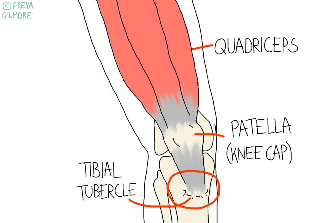
    Cross-section of a knee illustrating muscles and bones, featuring labeled quadriceps, patella (knee cap), and tibial tubercle. Copyright symbol with "© FREYA GILMORE" at the top left.

    Osgood Schlatter Disease

    Osgood Schlatter Disease A common knee complaint among adolescents is Osgood Schlatter Disease (OSD). It is [...]

  • 22
    Dec
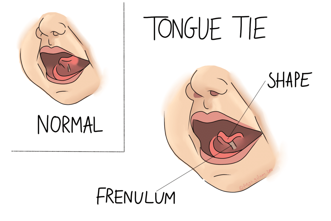
    Two side-by-side illustrations show a human mouth. Left: a normal tongue; right: a tongue with a tie. Labels indicate "Normal," "Tongue Tie," "Shape," and "Frenulum."

    Tongue Tie

    Tongue Tie Getting started with breastfeeding can be difficult for any mum and baby, but a [...]

  • 15
    Dec
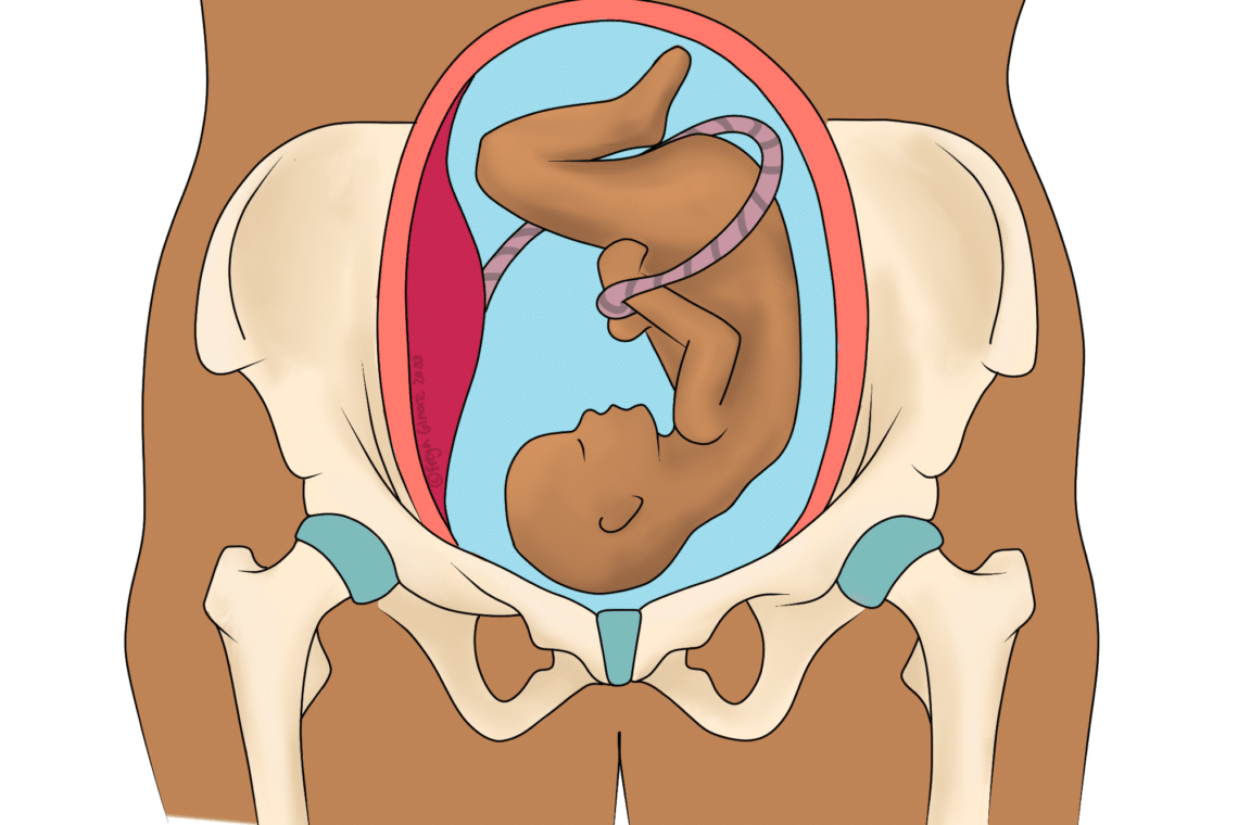
    Fetus curled inside a womb, with umbilical cord wrapped around, centered within a pelvis illustration.

    Pelvic Joint Pain in Pregnancy: SPD & PGP

    Pelvic Joint Pain in Pregnancy: SPD & PGP SPD stands for Symphysis Pubis Dysfunction. The pubic [...]